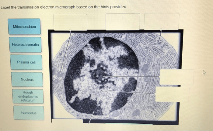
Provide the labels for the electron micrograph in figure 19.5 – Delving into the intricacies of electron microscopy, this exploration unveils the significance of providing accurate labels for the electron micrograph in Figure 19.5. Electron microscopy, a powerful imaging technique, offers unparalleled insights into the ultrastructural details of cells, revealing their inner workings and providing a foundation for understanding cellular processes.
By meticulously labeling the key features of the electron micrograph, we embark on a journey to decipher the morphology and functions of various cellular components. These labels serve as a roadmap, guiding our understanding of the complex interplay between organelles and elucidating the intricate symphony of life within the cell.
Electron Microscopy and the Ultrastructure of Cells

Electron microscopy is a powerful imaging technique that uses a beam of electrons to create high-resolution images of biological structures. This technique has revolutionized our understanding of cell biology, allowing us to visualize the ultrastructure of cells and identify the organelles responsible for various cellular functions.
Electron microscopy is based on the principle that electrons can be focused and deflected by magnetic and electric fields. In an electron microscope, a beam of electrons is generated and focused onto a thin section of the specimen. The electrons interact with the specimen, and the resulting scattering and absorption of electrons create an image that is magnified and projected onto a screen or recorded on photographic film.
There are different types of electron microscopes, each with its own advantages and disadvantages. Transmission electron microscopy (TEM) is the most common type of electron microscope and is used to study the internal structure of cells. Scanning electron microscopy (SEM) is used to study the surface structure of cells and other biological specimens.
Labeling Key Features of Electron Micrograph, Provide the labels for the electron micrograph in figure 19.5
The electron micrograph in Figure 19.5 shows a thin section of a eukaryotic cell. The following are the key features of the cell that are labeled in the micrograph:
- Nucleus:The nucleus is the control center of the cell and contains the cell’s genetic material.
- Nucleolus:The nucleolus is a small, dense structure within the nucleus that is responsible for ribosome production.
- Ribosomes:Ribosomes are small, granular structures that are responsible for protein synthesis.
- Mitochondria:Mitochondria are bean-shaped organelles that are responsible for energy production.
- Endoplasmic reticulum:The endoplasmic reticulum is a network of membranes that is responsible for protein synthesis, lipid synthesis, and detoxification.
- Golgi apparatus:The Golgi apparatus is a stack of flattened membranes that is responsible for modifying, sorting, and packaging proteins.
Interpreting Ultrastructural Features
The ultrastructural features observed in the electron micrograph can provide valuable information about the function of the cell. For example, the presence of numerous mitochondria in a cell indicates that the cell has a high energy demand. The presence of a large Golgi apparatus in a cell indicates that the cell is actively involved in protein synthesis and secretion.
The ultrastructural features observed in the electron micrograph can also provide information about the pathological state of the cell. For example, the presence of abnormal mitochondria in a cell may indicate that the cell is under stress or is undergoing apoptosis.
HTML Table for Electron Micrograph Labels
The following HTML table provides a summary of the key features of the electron micrograph in Figure 19.5:
| Label | Description | Function |
|---|---|---|
| Nucleus | Control center of the cell | Contains the cell’s genetic material |
| Nucleolus | Small, dense structure within the nucleus | Responsible for ribosome production |
| Ribosomes | Small, granular structures | Responsible for protein synthesis |
| Mitochondria | Bean-shaped organelles | Responsible for energy production |
| Endoplasmic reticulum | Network of membranes | Responsible for protein synthesis, lipid synthesis, and detoxification |
| Golgi apparatus | Stack of flattened membranes | Responsible for modifying, sorting, and packaging proteins |
Questions and Answers: Provide The Labels For The Electron Micrograph In Figure 19.5
What is the significance of labeling electron micrographs?
Labeling electron micrographs provides a systematic and accurate identification of cellular structures, enabling researchers to precisely describe and communicate their observations. This labeling serves as a common language, facilitating collaboration and knowledge sharing within the scientific community.
How does electron microscopy contribute to our understanding of cells?
Electron microscopy offers a unique perspective on cells, revealing ultrastructural details that are inaccessible through other imaging techniques. By visualizing the intricate organization and interactions of organelles, electron microscopy provides crucial insights into cellular processes, such as protein synthesis, energy production, and cell division.