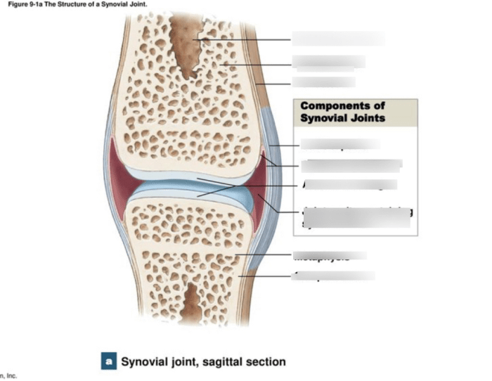
Art-labeling activity: structure of a typical synovial joint – Embark on an art-labeling activity that delves into the intricate structure of a typical synovial joint, an essential component of our musculoskeletal system. This exploration promises to illuminate the remarkable interplay of tissues and their functions, providing a comprehensive understanding of joint mechanics and movement.
As we embark on this journey, we will dissect each layer of the synovial joint, unraveling the unique properties and roles of articular cartilage, synovial membrane, synovial fluid, joint capsule, ligaments, tendons, and sensory receptors. Through this immersive experience, we will gain a profound appreciation for the complexity and resilience of the human body.
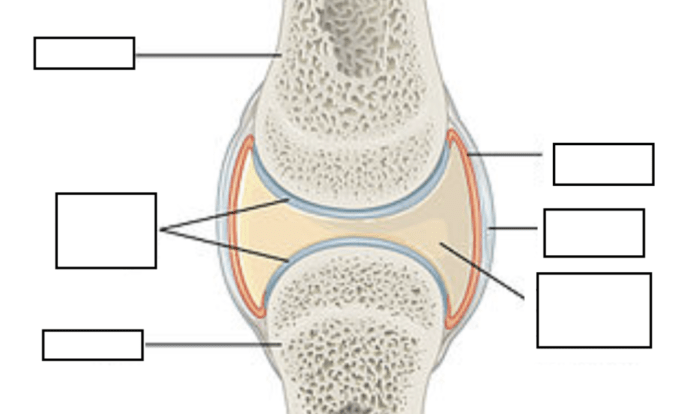
Synovial Joint Structure: Overview
Synovial joints are the most common type of joint in the human body. They are characterized by the presence of a synovial membrane, which produces synovial fluid that lubricates the joint and reduces friction during movement.
Structure
Function
Location
Articular cartilage
Covers the ends of bones and provides a smooth, gliding surface for joint movement.
At the ends of bones within the joint cavity
Synovial membrane
Lines the joint cavity and produces synovial fluid.
Inside the joint capsule
Synovial fluid
Lubricates the joint and reduces friction during movement.
Within the joint cavity
Joint capsule
Encloses the joint and provides stability.
Surrounds the joint cavity
Articular Cartilage: Function and Properties, Art-labeling activity: structure of a typical synovial joint
Articular cartilage is a specialized type of cartilage that covers the ends of bones within a synovial joint. It provides a smooth, gliding surface for joint movement and helps to distribute load across the joint.
Articular cartilage has unique properties that make it well-suited for its role in joint movement. It is elastic, allowing it to absorb shock and reduce stress on the joint. It also has a low coefficient of friction, which helps to reduce wear and tear on the joint during movement.
Synovial Membrane: Composition and Function
The synovial membrane is a thin layer of tissue that lines the joint cavity. It is composed of two layers: an inner layer of synovial cells and an outer layer of connective tissue.
The synovial membrane produces synovial fluid, which lubricates the joint and reduces friction during movement. Synovial fluid also contains nutrients that nourish the articular cartilage and other tissues within the joint.
Synovial Fluid: Characteristics and Importance
Synovial fluid is a viscous, clear fluid that fills the joint cavity. It is composed of water, proteins, and other nutrients.
Synovial fluid has several important functions, including:
Lubrication: Synovial fluid helps to reduce friction between the articular cartilage surfaces during joint movement.
Shock absorption: Synovial fluid helps to absorb shock and protect the joint from damage.
Nutrient transport: Synovial fluid contains nutrients that nourish the articular cartilage and other tissues within the joint.
Joint Capsule: Structure and Function
The joint capsule is a tough, fibrous membrane that surrounds the joint cavity. It is composed of two layers: an outer layer of dense connective tissue and an inner layer of synovial membrane.
The joint capsule provides stability to the joint and helps to prevent excessive movement. It also contains ligaments, which are bands of connective tissue that connect bones to each other and help to limit joint movement.
Ligaments and Tendons: Support and Stability
Ligaments are bands of connective tissue that connect bones to each other. They help to stabilize joints and prevent excessive movement.
Tendons are bands of connective tissue that connect muscles to bones. They help to transmit force from muscles to bones, allowing for joint movement.
Joint Movement and Biomechanics
Synovial joints allow for a wide range of movements, including flexion, extension, rotation, and abduction. The type of movement that is possible at a particular joint is determined by the shape of the joint and the ligaments that surround it.
The biomechanics of joint movement involves the interaction of muscles, tendons, and ligaments. Muscles generate force that is transmitted through tendons to bones, causing the bones to move. Ligaments help to control the range of movement and prevent excessive movement.
Joint Innervation and Sensory Receptors
Synovial joints are innervated by sensory receptors that provide information about the position and movement of the joint.
These sensory receptors include:
Proprioceptors: Proprioceptors are sensory receptors that provide information about the position of the joint.
Nociceptors: Nociceptors are sensory receptors that detect pain.
The information provided by these sensory receptors is essential for maintaining joint stability and preventing injury.
Joint Pathologies: Common Disorders and Injuries
Synovial joints are susceptible to a variety of disorders and injuries, including:
Osteoarthritis: Osteoarthritis is a degenerative joint disease that is characterized by the breakdown of articular cartilage.
Rheumatoid arthritis: Rheumatoid arthritis is an autoimmune disease that causes inflammation of the synovial membrane.
Sprains: Sprains are injuries to ligaments that can occur when a joint is twisted or turned awkwardly.
These are just a few of the many disorders and injuries that can affect synovial joints. Treatment for these conditions varies depending on the severity of the injury or disorder.
Query Resolution: Art-labeling Activity: Structure Of A Typical Synovial Joint
What is the primary function of articular cartilage?
Articular cartilage plays a crucial role in joint movement and load-bearing, providing a smooth, gliding surface that reduces friction and protects the underlying bone.
How does the synovial membrane contribute to joint function?
The synovial membrane produces synovial fluid, a viscous lubricant that nourishes and cushions the joint, facilitating smooth movement and reducing wear and tear.
What is the role of ligaments and tendons in joint stability?
Ligaments connect bones to bones, providing stability and limiting excessive movement, while tendons connect muscles to bones, enabling movement and transmitting force.